One of the questions that comes up most often when people first encounter scalar healing is some version of this: how could a field you cannot see or feel do anything meaningful to the body? It is a fair question, and it deserves a serious answer rather than a reassuring wave of the hand.
The answer begins not with scalar theory at all, but with something much more fundamental: the human body is already electric. It generates its own fields. It runs on electrical signals. It has been doing this, continuously, since before you were born. The question is not whether the body responds to electromagnetic phenomena. It clearly does. The more specific questions are which kinds of fields it responds to, through what mechanisms, and at what intensities. Those questions lead you from settled biology through emerging research and eventually to the open territory where scalar healing sits.
This article works through that progression carefully, separating what is well-established from what is still being investigated, and from what remains theoretical.
Part 1 — The Body's Own Electricity
Most people learn in school that the nervous system uses electrical signals, but the full picture of bioelectricity in the human body is considerably richer than that summary suggests. Electrical activity is not confined to neurons. It is a fundamental property of living cells at every level of biological organization.
Cell membrane potentials
Every cell in the human body maintains a voltage difference across its outer membrane. This voltage, called the resting membrane potential, arises because cells actively pump specific ions across their membranes using protein channels embedded in the membrane wall. Sodium and calcium ions are kept at higher concentrations outside the cell. Potassium ions are kept at higher concentrations inside. The result is a charge imbalance: the inside of most cells sits at approximately negative 70 millivolts relative to the outside [1].
Negative 70 millivolts sounds small, but across a membrane that is only about 7 nanometers thick, it represents an electric field strength of roughly 10 million volts per meter. Every resting cell in your body is maintaining what is, at the nanoscale, an extraordinarily intense electric field. This is not a passive side-effect of being alive. It is how cells regulate their internal chemistry, respond to signals from neighboring cells, and maintain the conditions necessary for their own function.
When that membrane potential is disrupted, when the voltage across the membrane changes significantly, the consequences are immediate and measurable. Cells that are stressed, injured, or cancerous typically show altered membrane potentials. The resting potential is, in a real sense, an indicator of cellular health [1].
Nerve signals
Neurons communicate by rapidly and temporarily reversing their membrane potential. When a neuron fires, sodium channels in the membrane open suddenly, allowing a rush of positively charged sodium ions into the cell. The interior voltage swings from negative 70 millivolts up to roughly positive 40 millivolts in about a millisecond. This electrical event, called an action potential, then propagates along the length of the neuron as each segment of membrane triggers the next [2].
The speed of propagation depends on the neuron’s diameter and whether it is coated in a fatty insulating layer called myelin. Myelinated neurons conduct signals at up to 120 meters per second. That is 432 kilometers per hour, fast enough for a nerve signal from your foot to reach your brainstem in under a hundredth of a second.
At the synapse, where one neuron meets the next, the electrical signal triggers the release of chemical neurotransmitters, which cross the gap and initiate a new electrical response in the receiving neuron. The brain, spinal cord, and peripheral nervous system are, in the most literal sense, an electrical network, generating and routing electrical signals continuously throughout every waking and sleeping moment of life [2].
The heart's electrical activity
The heart is driven by its own internal electrical pacemaker, a cluster of specialized cells in the upper right chamber called the sinoatrial node. This node generates spontaneous electrical impulses at a resting rate of 60 to 100 times per minute, each impulse spreading across the heart muscle in a precise sequence that coordinates the contraction of the four chambers. The electrical wave travels through the atria first, then pauses briefly at the atrioventricular node before descending through the ventricles along specialized conducting pathways [3].
The electrocardiogram, or ECG, records the electrical activity generated by this process from electrodes placed on the skin. The peaks and troughs of the ECG trace correspond to specific electrical events in the cardiac cycle and can reveal a remarkable amount about the health and behavior of the heart. The fact that this electrical activity is detectable on the skin’s surface illustrates something important: the body’s internal electrical fields are not contained within individual cells. They propagate outward through surrounding tissue and generate measurable fields outside the body.
The heart generates the strongest electromagnetic field of any organ in the body. Its electrical field extends several meters beyond the skin surface. Its magnetic field, while far weaker, is measurable with sensitive magnetometers at a distance of several centimeters. This is not fringe science. Magnetocardiography, the recording of the heart’s magnetic field, is an established clinical diagnostic technique used in specialized medical centers [3].
Part 2 — How External Fields Interact with the Body
Given that the body runs on electrical signals, the next question follows naturally: can fields from outside the body influence the body’s own electrical activity? The answer, supported by decades of research, is yes. The more specific and contested question is under what conditions, at what intensities, and through what mechanisms.
Mechanotransduction and electrosensing at the cellular level
Cells detect and respond to external mechanical and electromagnetic stimuli through specialized proteins embedded in their membranes. Some of these proteins are mechanosensitive ion channels: they open or close in response to mechanical deformation of the membrane, allowing ions to flow and changing the cell’s electrical state. Others are voltage-sensitive channels: they open or close in response to changes in the local electric field, cascading into downstream signaling events [4].
This cellular electrosensitivity is not a marginal phenomenon. It is central to how tissues grow, repair themselves, and maintain their organization. Applied electric fields are known to influence the direction of cell migration during wound healing. A cut in the skin generates a skin battery effect, an electrical current that flows from the edges of the wound toward its center, which helps guide cells to close the wound. Disrupting this current disrupts wound healing. Restoring or augmenting it, using externally applied electric fields, can accelerate repair [4].
PEMF and established clinical applications
The clearest demonstration that external electromagnetic fields can produce meaningful biological effects comes from pulsed electromagnetic field therapy, PEMF. As covered in earlier articles in this series, PEMF received its first FDA approval in 1979 for the treatment of bone fractures that had failed to heal through normal biological processes. The mechanism involves electromagnetic induction: a pulsing magnetic field generates small electrical currents within the tissue, and those currents appear to stimulate the activity of bone-forming cells called osteoblasts [5].
Multiple subsequent FDA clearances have followed, and the research literature on PEMF now spans several decades and covers applications from osteoarthritis pain management to peripheral nerve regeneration to transcranial magnetic stimulation for treatment-resistant depression. These are mainstream medical technologies, used in hospitals and clinics, with controlled clinical trial evidence behind them. They establish, definitively, that external electromagnetic fields can produce specific, reproducible, clinically meaningful changes in biological tissue [5].
The intensities involved in PEMF therapy are modest by engineering standards. The magnetic fields used in clinical PEMF devices are typically in the range of a few gauss, far weaker than the fields produced by a standard MRI machine. The induced currents in tissue are in the microampere range. The body’s response to fields this weak, producing effects measurable in clinical outcomes, tells you something important about the sensitivity of biological systems to electromagnetic stimulation.
Transcranial magnetic stimulation and the brain
Transcranial magnetic stimulation, TMS, applies brief, intense magnetic pulses to specific regions of the skull, inducing electrical currents in the brain tissue beneath. Repeated TMS sessions have been shown in controlled clinical trials to reduce symptoms of major depression in patients who have not responded to medication, leading to FDA approval for this application [6]. More recent research has explored TMS for post-traumatic stress disorder, obsessive-compulsive disorder, and chronic pain.
TMS works not by heating tissue or causing structural changes, but by modulating the electrical activity of neurons through electromagnetic induction. The neurons respond to the externally induced electrical currents exactly as they would respond to signals arriving through their normal biological pathways. From the neuron’s perspective, an externally induced electrical signal is an electrical signal. The body does not distinguish the source.
Part 3 — Emerging Research: Biophotons and Bioelectric Patterning
Beyond the established mechanisms above, a number of research areas are generating findings that extend our picture of bioelectricity into new territory. These are not yet established to the same standard as PEMF or TMS, but they are being investigated by credentialled scientists with published results that merit attention.
Biophotons and cellular light signaling
Living cells emit ultra-weak light, a phenomenon called biophoton emission, first described systematically by Alexander Gurwitsch in the 1920s and later rigorously documented by Fritz-Albert Popp and others from the 1970s onward. The emission is real and is now accepted by mainstream biology as a confirmed phenomenon, arising from oxidative metabolic processes within the cell [7].
What remains open is Popp’s further hypothesis: that biophotons are not merely a byproduct of metabolism but a coherent signaling medium that cells use to coordinate activity across tissue. If cells communicate through coherent light, that would represent an electromagnetic communication channel operating at visible light frequencies, entirely within the body, that conventional biology has not yet integrated into its models. Several research groups are actively investigating this. No consensus has been reached, but the hypothesis has not been disproven [7].
Bioelectric patterning and developmental biology
One of the more striking recent developments in biology is the growing recognition that bioelectric signals, patterns of voltage across tissues and organs, play an instructive role in development, not just a permissive one. Michael Levin’s laboratory at Tufts University has demonstrated that bioelectric patterns in developing embryos carry positional information: they tell cells where they are in the body and what kind of tissue they should become. Disrupting these bioelectric patterns produces developmental abnormalities. Restoring or altering them can redirect tissue fate [8].
This is a significant shift in how developmental biologists think about the body. The genetic code specifies the molecular machinery. The bioelectric pattern specifies how that machinery is organized in space. Both layers of information appear to be necessary. And the bioelectric layer, because it is electromagnetic in nature, is in principle responsive to external electromagnetic fields in ways that the genetic layer is not.
Levin’s group has shown that it is possible to induce the growth of an eye in a non-eye location of a frog embryo by manipulating bioelectric signals alone, without modifying any genes. They have also demonstrated regeneration of tissue in non-regenerating species by manipulating bioelectric patterns. This research is peer-reviewed, reproducible, and represents a genuine expansion of what biology understands about the role of electromagnetic fields in living systems [8].
Part 4 — The Evidence Gap: From Established Bioelectricity to Scalar Healing
The established science reviewed above demonstrates something important and specific: the human body is an electromagnetic system, sensitive to external electromagnetic fields, with biological processes that can be meaningfully influenced by appropriately designed electromagnetic stimulation. That is not a small finding. It is the foundation that makes the entire conversation about field-based healing scientifically coherent rather than arbitrarily speculative.
But it does not, on its own, validate the specific claims of scalar healing. Getting from established bioelectricity to the claims made for scalar fields requires crossing a gap, and it is worth being honest about exactly where that gap lies and how wide it is.
The mechanism gap
PEMF, TMS, and the bioelectric patterning research all work through mechanisms that are understood well enough to design experiments, make predictions, and test results. The magnetic field induces electrical currents through Faraday’s law. The currents activate voltage-gated ion channels. The ion channel activity produces downstream biological responses. Each link in the chain can be measured, disrupted, and restored experimentally.
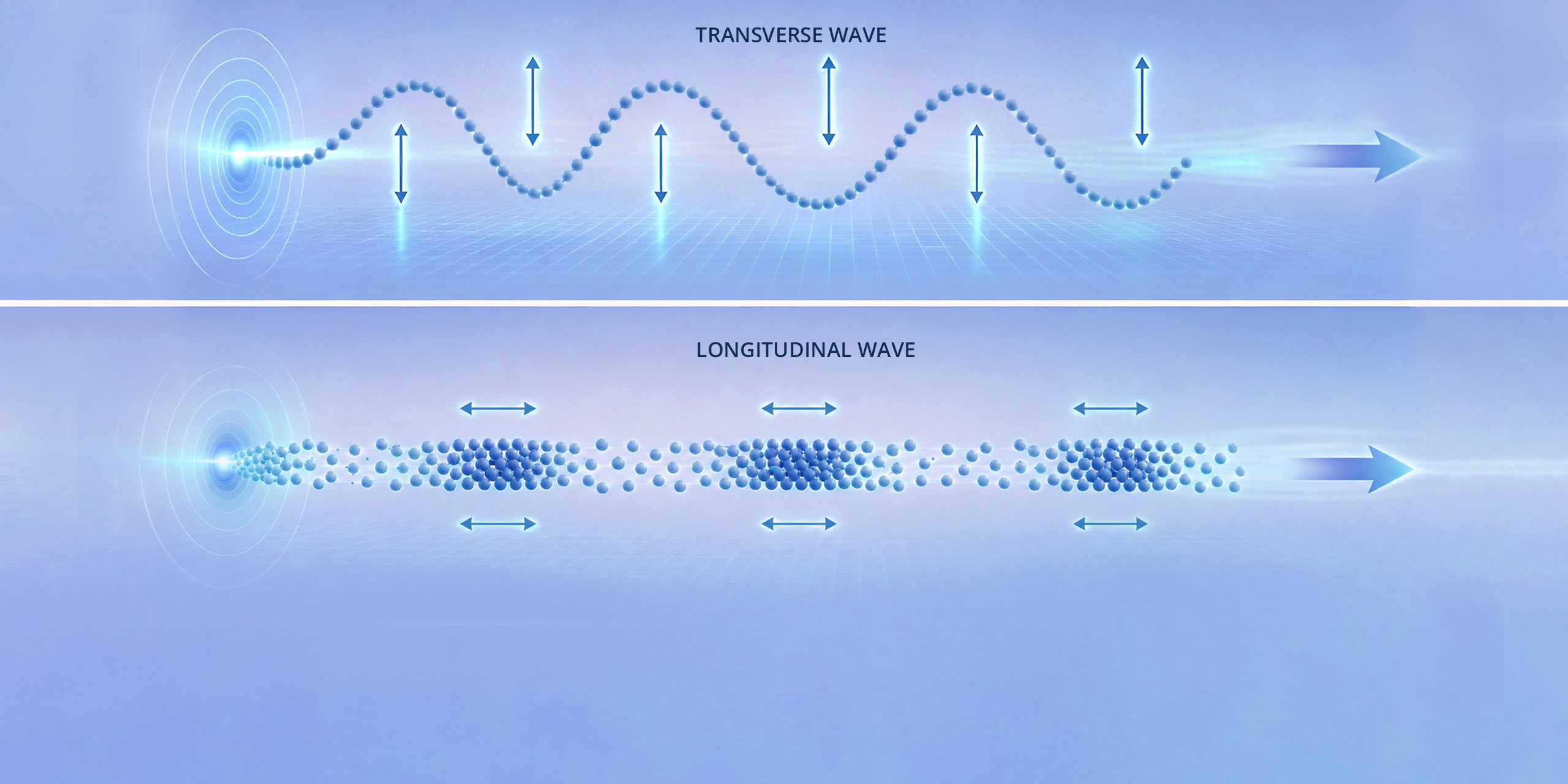
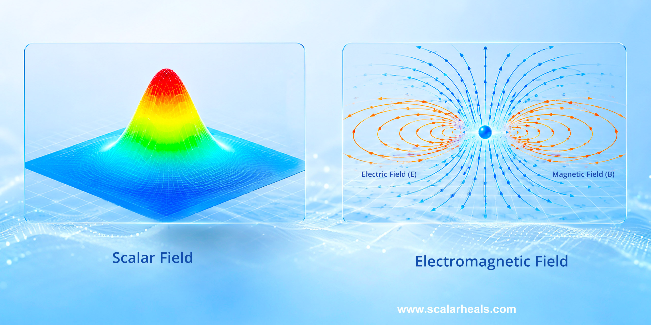
For scalar healing, the proposed mechanism involves the scalar potential component of the electromagnetic field, possibly including longitudinal wave propagation of the kind Meyl describes, interacting with the body’s bioelectric systems in ways that conventional field-based therapies do not. This is a coherent theoretical proposal. The problem is that the mechanism has not been characterized at the level of the ion channel, the cell membrane, or the tissue. We do not yet know which biological structures would detect and respond to scalar potential components specifically, as opposed to the conventional electric and magnetic field components that the same devices might also be generating [9].
The measurement gap
A related problem is measurement. PEMF researchers can characterize exactly what their devices produce: magnetic field strength, pulse frequency, waveform, and induced current density in tissue. Those parameters can be related to biological outcomes in a quantitative way. For scalar devices, the field produced is described in terms that are not yet measurable by instruments that have been independently calibrated and validated. This makes it difficult to establish dose-response relationships, compare devices, or design controlled trials that would clearly attribute any observed effect to the scalar component rather than to the conventional electromagnetic components that may be present simultaneously [9].
The clinical evidence gap
Perhaps most straightforwardly, the clinical trial evidence for scalar healing specifically is not yet at the level that the established therapies have reached. PEMF has randomized controlled trials, FDA approvals, and meta-analyses. TMS has the same. Scalar healing has user reports, practitioner observations, and a small number of case studies. These are real forms of evidence, and they are consistent enough across a large enough community that they are not easily dismissed. But they are not the same thing as controlled trials, and the difference matters when trying to determine whether an effect is real and what is causing it [9].
Part 5 — What the Evidence Suggests, Taken Together
The picture that emerges from reviewing all of this is more encouraging for scalar healing than the simple ‘no evidence’ dismissal that some skeptics offer, and less settled than the confident clinical language that some practitioners use.
The body is genuinely sensitive to external electromagnetic fields. That sensitivity operates through documented biological mechanisms at the cell membrane, the ion channel, and the tissue level. The sensitivity extends to fields that are far weaker than most people would assume, as PEMF’s clinical effectiveness at modest field intensities demonstrates. The role of bioelectric patterning in development and healing is emerging as a significant field in mainstream biology, opening new ways of thinking about how electromagnetic information is organized and used in living systems.
Against that backdrop, the idea that scalar field devices might influence the body’s bioelectric systems through mechanisms not yet fully characterized is not implausible. It is an extrapolation from a real and increasingly well-understood biological foundation. The extrapolation may be correct. It has not yet been validated through the kind of mechanistic and clinical research that would establish it to the standard that PEMF and TMS now meet.
The body is not a passive object that electromagnetic fields act upon from outside. It is itself an electromagnetic system, generating fields, communicating through them, and organized by them from its earliest development. Understanding that is the first step toward understanding why field-based approaches to health are not inherently implausible, and why the specific mechanisms that scalar healing proposes deserve serious rather than dismissive investigation.
Resources:
- [1] Alberts, B., Johnson, A., Lewis, J., Raff, M., Roberts, K., & Walter, P. (2014). Molecular Biology of the Cell (6th ed.). Garland Science. [Comprehensive reference on cell membrane potentials, ion channels, and the biophysics of cellular electrical activity]
- [2] Kandel, E. R., Schwartz, J. H., Jessell, T. M., Siegelbaum, S. A., & Hudspeth, A. J. (2012). Principles of Neural Science (5th ed.). McGraw-Hill. [Standard neuroscience reference covering action potentials, signal propagation, and synaptic transmission]
- [3] Guyton, A. C., & Hall, J. E. (2020). Textbook of Medical Physiology (14th ed.). Elsevier. [Standard medical physiology reference covering cardiac electrical activity, the ECG, and the heart’s electromagnetic field]
- [4] Jaffe, L. F., & Vanable, J. W. (1984). Electric fields and wound healing. Clinics in Dermatology, 2(3), 34-44. https://doi.org/10.1016/0738-081X(84)90025-0 [Classic paper on the skin battery effect and the role of endogenous electric fields in wound healing]
- [5] Pilla, A. A. (2013). Nonthermal electromagnetic fields: From first messenger to therapeutic applications. Electromagnetic Biology and Medicine, 32(2), 123-136. https://doi.org/10.3109/15368378.2013.776335
- [6] Lefaucheur, J. P., et al. (2014). Evidence-based guidelines on the therapeutic use of repetitive transcranial magnetic stimulation. Clinical Neurophysiology, 125(11), 2150-2206. https://doi.org/10.1016/j.clinph.2014.05.021
- [7] Popp, F. A., & Beloussov, L. (Eds.). (2003). Integrative Biophysics: Biophotonics. Springer. https://doi.org/10.1007/978-94-017-0373-4
- [8] Levin, M. (2012). Morphogenetic fields in embryogenesis, regeneration, and cancer: Non-local control of complex patterning. Biosystems, 109(3), 243-261. https://doi.org/10.1016/j.biosystems.2012.04.005
- [9] Oschman, J. L. (2000). Energy Medicine: The Scientific Basis. Churchill Livingstone. [Overview of bioelectromagnetic field effects in living systems and the theoretical basis for field-based therapeutic approaches]